Iba1 is a calcium-binding protein with a molecular weight of 17,000 specifically expressed in macrophage/microglia. Recently, microglia has attracted attentions because neurological damage effect by production of NO, TNF-α, and IL-1β have been proved in addition to its role in neurotrophic effects/neuroprotective actions. These products are antibodies that specifically recognize Iba1, and are available for a microglial marker.
In particular, "Anti Iba1, Rabbit (for immunocytochemistry) Code No. 019-19741" is used by many worldwide researchers as a standard for microglial marker antibody. There are also many references in major journals such as Nature, Cell etc.
References
Anti-Iba1, rabbit (for immunocytochemistry) product code: 019-19741
Search with google scholar with the keyword "Iba1 019-19741 Wako"
For detailed literature information, please refer to HERE.
| Journal title | The number of references | IF in 2017/2018 |
|---|---|---|
| Nature | 16 | 41.577 |
| Cell | 6 | 31.398 |
| Nature Medicine | 5 | 32.621 |
| Nature Neuroscience | 23 | 19.912 |
| Nature Immunology | 3 | 21.809 |
| Nature Biotechnology | 2 | 35.724 |
| Nature Methods | 2 | 26.919 |
| Neuron | 11 | 14.318 |
| Total | 68 |
Application 1
Application data for immunohistochemistry, rat frozen section
Experimental condition
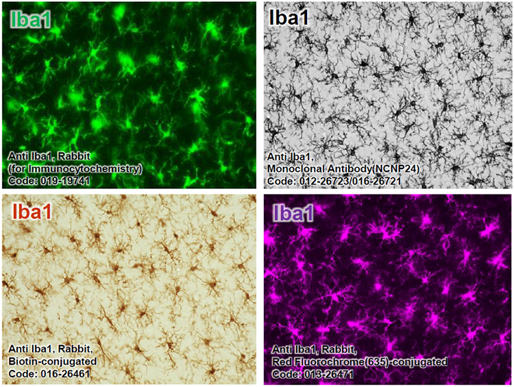
Code No. 019-19741/012-26723/016-26721
Tissue: Rat brain cortex
Section: Frozen section
Antibody concentration: 1/1,000
Code No. 016-26461/013-26471
Tissue: Rat brain cortex
Section: Frozen section
Antibody concentration: 1/200
These data were provided by Sanagi,T., Manabe,T., Ichinohe, N., and Kohsaka, S., National Center of Neurology and Psychiatry in Japan.
Application 2
Application Data –immunohistochemistry paraffin section-
016-27691:Anti Iba1, Rabbit(for Paraffin Section)
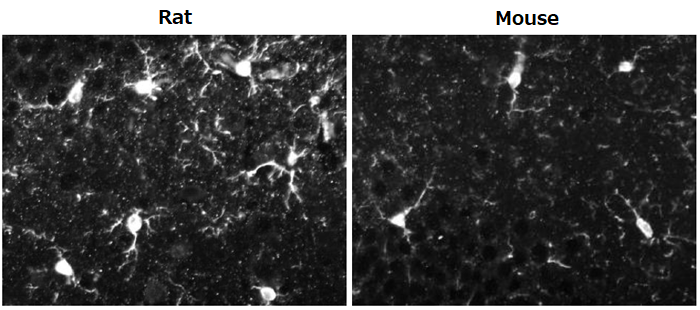
Experimental condition
Tissue: mouse and rat hippocampus neighborhood
Section: paraffin section
Antibody concentration: 1/1,000
Application 3
Application Data -western blotting-
016-20001: Anti Iba1, Rabbit (for Western Blotting)
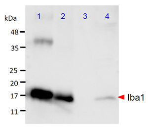
Lane
- Iba1 protein (10 ng)
- Rat microglia (10 µg)
- Rat neuron (10 µg)
- Rat cortex (150 µg)
Experimental condition
SDS-PAGE: 5.5 % stacking gel, 12.5 % running gel, 100V
Blocking: 3 % skim milk in TBS at RT for 1h
Primary antibodies:anti-Iba1 antibody(1/1000) in 3 % skim milk in TTBS at 4 °C O/N
Secondary antibody: peroxidase labeled anti-rabbit antibody (1/5000) in 3 % skim milk in TTBS at RT for 1h
The data was provided by Sanagi,T., Ichinohe, N., and Kohsaka, S., National Center of Neurology and Psychiatry in Japan.
Antibody Information
| Product Name |
Polyclonal antibody | Monoclonal antibody | |||||
|---|---|---|---|---|---|---|---|
| Anti Iba1, Rabbit (for Immuno- cytochemistry) |
Anti Iba1, Rabbit (for Paraffin Section) |
Anti Iba1, Rabbit Biotin- conjugated |
Anti Iba1, Rabbit Red Fluorochrome (635)- conjugated |
Anti Iba1, Rabbit (for Western Blotting) |
Anti Iba1, Monoclonal Antibody (NCNP24) |
Anti Human Iba1, Monoclonal Antibody (NCNP27) |
|
| Cat Code (Pkg. Size) |
019-19741 (50 µg) |
013-27691 (50 µg) |
016-26461 (100 µL) |
013-26471 (100 µL) |
016-20001 (50 µg) |
012-26723 (10 µL) 016-26721 (50 µL) |
017-27591 (10 µL) 013-27593 (50 µL) |
| Antigen | Synthetic peptide (C-terminal of Iba1) |
Synthetic peptide (C-terminal of Iba1) |
|||||
| Clone No. | - (Polyclonal) |
NCNP24 | NCNP27 | ||||
| Subclass | Rabbit IgG | Mouse IgG1 | Mouse IgG2 | ||||
| Conjugate | - | - | Biotin | Red Fluorochrome (Ex=634, Em=654 nm) |
- | - | - |
| Concentration | 0.5 mg/mL | 0.5 mg/mL | 0.5 mg/mL | 0.5 mg/mL | 0.5 mg/mL | 1.3 mg/mL (First lot) |
1.0 mg/mL (First lot) |
| Buffer | TBS | TBS | PBS, 0.05% NaN3 | PBS, 0.05% NaN3 | TBS | 50% Glycerol / TBS, 0.05% NaN3 | 50% Glycerol / TBS, 0.05% NaN3 |
| Cross- reactivity |
Mouse, rat, human,etc※ | Mouse, rat | Mouse, rat | Mouse, rat | Mouse, rat, human | Mouse, rat, marmoset | Human |
| Applications | ■Immunohisto- chemistry (frozen section) 1:500-1,000 ■Immunohisto- chemistry 1:500-1,000 |
■Immunohisto- chemistry (paraffin section) 1:500-1,000 |
■Immunohistochemistry (frozen section) 1:200-2,000 |
■Western Blotting 1:500-1,000 |
■Immuno- histochemistry (frozen section, DAB) 1:500-2,000 ■Immuno- histochemistry (frozen section, fluorescent) 1:100 |
■Immuno- histochemistry (paraffin section, DAB) 1:100-1,000 |
|
| Reference | 1)2)3)4)5) | - | 6) | - | 7)8) | 9)10) | - |
- The paper using samples of dog1), cat2), pig3), marmoset4), and zebrafish5)
References
- Ahn, J.H., et al.: Lab. Anim. Res., 28, 3, 165 (2012).
- Ide, T., et al.: J. Vet. Med .Sci., 72, 1, 99 (2010).
- Gaige, S., et al.: Neurotoxicology., 34, 135(2013).
- Rodriguez-Callejas, J.D. et al.: Front. Aging Neurosci., 8, 315(2016).
- Fantin, A., et al.: Blood, 116, 5, 829 (2010).
- Jones, M. E., et al.: Brain Behav. Immun., 67, 355(2018).
- Sun, J.S. et al.: Mol. Med. Rep., 12, 2, 2677(2015).
- Grishchuk, Y., et al.: Am. J. Pathol., 186, 1, 199(2016).
- Wan, S., et al.: J. Neuroinflammation, 15, 31(2018).
- Chen, Y., et al.: Annals of Clinical and Translational Neurology,10.1002/acn3.513 (2017).
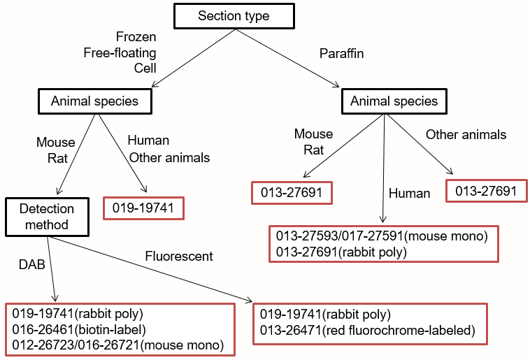
Select Flowchart